 (standing),
oblique,
(standing),
oblique, 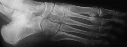 Harris
views. Tarsal coalition of calcaneonavicular appears to be present. The
subtalar joint does not appear to be involved.
Harris
views. Tarsal coalition of calcaneonavicular appears to be present. The
subtalar joint does not appear to be involved.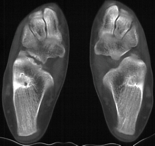 CT scan - Confirms the presence of a calcaneonavicular fibrous or
cartilaginous bar.
CT scan - Confirms the presence of a calcaneonavicular fibrous or
cartilaginous bar. JOHN ERGENER, M.D., Orthopaedic Resident
November 14,1995
CLINICAL CASE PRESENTATION
ORTHOPAEDIC DEPARTMENT
THE ALFRED I. DUPONT INSTITUTE
WILMINGTON, DELAWARE
(standing),
oblique, Harris
views. Tarsal coalition of calcaneonavicular appears to be present. The
subtalar joint does not appear to be involved.
CT scan - Confirms the presence of a calcaneonavicular fibrous or
cartilaginous bar. TREATMENT: Conservative measures of shoe modification and activity restriction
were instituted about a year ago. There was little resolution of the symptoms.
An additional attempt was madewith immobilization in a short leg walking
cast for approximately six weeks. The pain resolved while in the cast but
rapidly returned once the cast was removed. At this point surgical intervention
was performed. The calcaneonavicular coalition 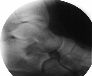 was excised with interposition of the extensor digitorum brevi. Significant
subtalar motion was regained. The patient was placed in a short leg cast
for 4 weeks. The cast was then removed and an AFO was fashioned. The patient
was gradually advance to weight bearing as tolerated. Subtalar mobilization
was encouraged. She has had resolution of her symptoms up to this point.
was excised with interposition of the extensor digitorum brevi. Significant
subtalar motion was regained. The patient was placed in a short leg cast
for 4 weeks. The cast was then removed and an AFO was fashioned. The patient
was gradually advance to weight bearing as tolerated. Subtalar mobilization
was encouraged. She has had resolution of her symptoms up to this point.
Connection of two or more tarsal bones. This bridge may be bony, cartilaginous or fibrous.
Estimated to be less than 1%. Talocalcaneal and calcaneonavicular are the most common. The incidence of bilaterality is approximately 50 %.
Unclear. It appears to be a defect in segmentation and differentiation of the primitive mesenchyme. Present in embryo confirming congenital rather than developmental origin. Proposed autosomal dominant inheritance with variable penetrance.
Most patient become symptomatic at the time w hen the coalition begins to ossify. About 3-5 years of age f or talonavicular coalitions (rare), 8-12 years for calcaneonavicular coalitions and 12-16 years for talocalcaneal coalitions. The pain is usually gradual in onset. Patients may complain of multiple sprains. This may be due to the increased stiffness of the subtalar joint leading to abnormal stresses thru the ankle. Walking on uneven ground may prove to be problematic. Some adults may become symptomatic after a traumatic event. The pain is worse with activity and usually gets better with rest. However, as the process continues many individuals become more limited in their activities.
Usually reveals tenderness at the location of the coalition. The medial aspect of the foot just anterior to the medial malleolus for a talocalcaneal coalition. This reflects that most talocalcaneal bridges involve the medical facet. The calcaneonavicular bar locates to the anterolateral aspect of the foot in the region of the sinus tarsi. Pes planus and heel valgus may b e present. A misnomer of peroneal spastic flatfoot has been used to describe the flat foot associated with tarsal coalitions. True muscle spasm is not present according to EMG studies. Rather, the peroneal muscles are shortened and attempted foot inversion causes pain b y stretching these muscles. Subtalar motion is decreased. The ankle joint may have increased laxity and give the false impression of greater subtalar motion.
Radiographs - AP, lateral, oblique at 45 degrees, and Harris view. A coalition may be difficult to pick up on the AP and lateral views due to the overlap of bones but there may be some subtle clues. Beaking of the talus or narrowing of the subtalar joint may be present. Calcaneonavicular bony bridges can be seen on the lateral view with the classic "anteater nose" coming from the calcaneous. Harris view may be helpful to evaluate the subtalar joint but a CT is often obtained to rule out subtalar coalition. The ankle may remodel to a ball and socket joint secondary to limited subtalar motion. Coronal CT cuts are most helpful in evaluating talocalcaneal bony bridges while transverse cuts are used for calcaneonavicular bars.
Since many adults with tarsal coalition are asymptomatic, the first line of treatment is conservative measures. Shoe modification and change in activity are attempted. An effort to stabilize the heel is made to decrease the stresses across a stiff subtalar oint. If this fails, a period of immobilization (3-6 weeks) may be attempted using a short leg walking cast. Reportedly up t a third of patients respond to non-operative intervention.
If conservative treatment fails it is necessary to intervene surgically. Significant relief can be obtained by resection of the coalition. It is, however, necessary to place a layer of material in the space to avoid a recurrence In the case of a calcaneonavicular coalition, the bar is resected and the extensor digitorum brevis is interposed.
The talocalcaneal coalition can be more problematic. Resecting a large amount of the medial facet can cause a disturbance of the weight bearing relationship of the foot. The coalition may be taken down if in fact it is not too large and there is not significant degenerative changes. Interposition of fat will prevent reformation. If there is significant degenerative changes, then a triple arthrodesis serves these individuals well. However, it should not be done until late adolescence or adulthood.