SUBACUTE EPIPHYSEAL OSTEOMYELITIS
TIMOTHY P. DOMER, D.O., Orthopaedic Surgery Resident
KIRK DABNEY M.D., Attending Pediatric Orthopaedic Surgeon
January, 29, 1996
CLINICAL CASE PRESENTATION
ORTHOPAEDIC DEPARTMENT
THE ALFRED I. DUPONT INSTITUTE
WILMINGTON, DELAWARE
CASE HISTORY:
PATIENT 1.
- HISTORY: This 9-year-old female who presented to the emergency room
with a four day history of mild right knee pain. She limps on the affected
extremity when ambulating but has minimal to no pain at rest. No history
of trauma was reported. She has had mildly elevated temperatures over the
past several days according to the mother. There is no history of joint
stiffness or chronic fatigue. No other joints are involved. There is no
history of tic exposure. She has been otherwise healthy in the past.
- PHYSICAL EXAM: On physical examination of the right knee there is no
limitation in flexion or extension. There is mild diffuse tenderness that
is poorly localized over the medial femoral condyle. There is no soft tissue
swelling or joint effusion noted. No pain is elicited with ROM of the knee.
The remaining extremities are without tenderness or swelling. Temperature
is 38.8* C
- LABS: WBC 13,400 Differential-- 73% polys, 8% lymphs, 1% bands, Sed
rate 41mm/hr
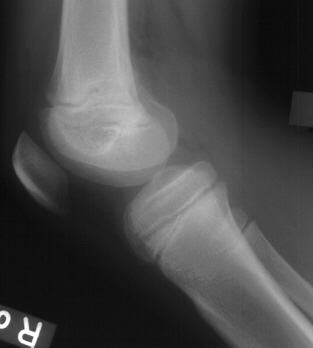
- XRAYS: No bony abnormalities noted on plain x-ray of the knee


- TREATMENT: The patient was discharged home and re-checked the following
day. Because of persistent tenderness over the knee and new physical findings
of swelling and pain with ROM of the knee, the following diagnostic studies
were obtained:
- Bone scan: Revealed increased uptake in the region of
the distal femur with a predominant epiphyseal location and slight uptake
in the metaphysis on the flow and pool images
 .
Delayed images showed focally increased uptake in the epiphysis of the
distal right femur
.
Delayed images showed focally increased uptake in the epiphysis of the
distal right femur .
.
- CT scan: No subperiosteal fluid collections were noted.
There was an area in the region of the distal femoral epiphysis that contained
a difference in density from the surrounding bone.
- Ultrasound: Soft tissue swelling in the region of the
distal femur
The patient was taken to the operating room for fluoroscopic guided
needle aspiration of the distal femoral metaphysis and epiphysis and the
proximal tibial metaphysis. Aspiration of the knee joint was also performed.
Final cultures: Blood cultures obtained at initial presentation
were positive for Staphylococcus aureus. Aspirates of the distal femoral
epiphysis were positive for Staphylococcus aureus
- FOLLOW-UP: The patient remained in the hospital for a total of 14 days.
She was discharged on IV cefuroxime to complete a six week course of antibiotic
therapy. At seventeen months follow-up she had painless full ROM of her
knee and no radiographic evidence of bony abnormality.
SUBACUTE EPIPHYSEAL OSTEOMYELITIS
Pathogenesis:
Predisposing factors that may influence the septic process in bone:
- Host resistance
- Virulence of infecting organism
- Adequacy of antibiotic therapy
- Trauma resulting in vascular injury and hypoxemia in bone
Staphylococcus aureus is the most common bacterial isolate, and it has
been found that S. aureus has a certain affinity for epiphyseal cartilage.
Pathoanatomy:
- Main blood supply to the epiphyseal end of long bones is through the
Hunter circle -- a large encircling artery that gives off branches to both
the metaphysis and epiphysis.
- Arcades are formed from epiphyseal branches which eventually end in
venous sinusoids in the form of loops, which may be analogous to the sluggish
blood flow that occurs in the metaphysis.
- The blood supply in the venous loops may be analogous to the sluggish
flow in the metaphyseal sinusoids, which is often the site of acute hematogenous
osteomyelitis.
- Physeal blood supply peripherally is via epiphyseal and periosteal
circulation, whereas central circulation is solely provided by epiphyseal
circulation.
Radiographic features:
- Benign appearing-- lesion is lytic, circular, and well circumscribed
with a sclerotic margin
- Malignant appearing-- bony cortex is eroded
Radiographic differential diagnosis (of all locations of subacute osteomyelitis)
- chondroblastoma
- osteoid osteoma
- Ewing's sarcoma
- eosinophilic granuloma
- unicameral bone cyst
- Brodies abscess
- osteogenic sarcoma
Radiographic classification:
Ia-- metaphyseal; punched out lucency suggestive of eosinophilic granuloma
IIb-- metaphyseal ; sclerotic margin with classic appearance for Brodies
abscess
III-- diaphyseal ; localized cortical and periosteal reaction simulating
osteoid osteoma
IV-- diaphyseal ; onion-skin periosteal reaction simulating Ewing's
sarcoma
V-- epiphyseal ; concentric radiolucency
VI-- vertebral body ; erosive or destructive process
Treatment:
Ross and Cole used the radiographic appearance of lesions associated
with subacute osteomyelitis to classify their patients into two groups
as follows:
- Group 1 -- Aggressive lesions- the initial presentation of these children
was that of primary malignant bone tumors based on clinical, radiographic
and hematologic studies.
- Group 2 -- Cavities in the region of the metaphysis and epiphysis-
these children presented with a more typical picture of subacute osteomyelitis
having cavities involving either the metaphysis or the epiphysis or both.
All patients were initially treated with two days of intravenous antibiotics
and then were switched to oral antibiotics to complete a six week course.
Results:
Treatment of group 1 lesions
- Consisted of curettage followed by six weeks of antibiotics and immobilization
-- 25/26 healed.
Treatment of group 2 lesions
- Those not operated on at the time of presentation -- 32/37 healed
without operation, including all 5 epiphyseal lesions. Of those not healing
initially without operation, three were confined to the metaphysis and
two were combined metaphyseal and epiphyseal lesions. All of these went
on to heal after curettage and an additional six week course of antibiotics.
- Those operated on at the time of presentation -- 7/11 patients underwent
drainage and eventually healed after antibiotics; 4/11 patients had biopsies
to establish the diagnosis of subacute osteomyelitis- there is no mention
of formal drainage or whether there was complete healing.
References:
- Green,N.E., Beauchamp,R.D., Griffin, P.P. Primary subacute epiphyseal
osteomyelitis. J Bone Joint Surg 63-A:107, 1981.
- Green, N.E. Osteomyelitis of the epiphysis. In Behavior of the growth
plate. Raven press, 1988.
- Letts, R.M. Subacute osteomyelitis of the growth plate. In Behavior
of the growth plate. Raven press, 1988.
- Lindenbaum,S. Alexander, H. Infections stimulating bone tumors: a review
of subacute osteomyelitis. Clin. Orthop. 184:193, 1984.
- Roberts, J.M., Drummond, D.S., Breed, A.L., Chesney, J. Subacute hematogenous
osteomyelitis in children: a retrospective study. J Pediatr Orthop 2:249,
1982.
- Ross, E.R.S., Cole, W.G. Treatment of subacute osteomyelitis in childhood.
J Bone Joint Surg 67-B:443, 1985.