 ,
12 yrs
,
12 yrs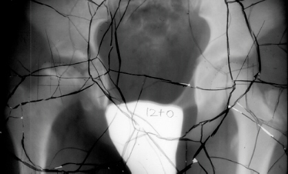
 ,
17 yrs
,
17 yrsMASAFUMI HOMMA, M.D.,D.M.Sc, Research Fellow of Orthopaedic Surgery
J. RICHARD BOWEN, M.D., Chairman of Pediatric Orthopaedic Surgery
April. 9,1996
CLINICAL CASE PRESENTATION
ORTHOPAEDIC DEPARTMENT
THE ALFRED I. DUPONT INSTITUTE
WILMINGTON, DELAWARE
F.T. is a 44 y. male. Perthes disease on the right hip.
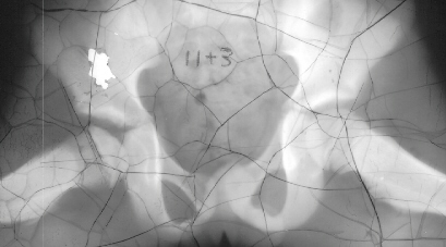
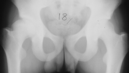
The parents first noted at the time the patient was 10y and 9 m old that he would limp on the right side after a full day of activity. He also started to have a occasional hip pain. Since the hip pain became progressively worse, he visited local clinics and was referred to AIDI at the age of 11y and 3 m. At the first examination abduction was 40 degrees bilaterally but internal rotation was limited to 0 degree on the right. The leg lengths were equal and no Trendelenburg sign was observed. X-ray showed the decreased epiphyseal height and medial joint opening. Snyder sling was started. At 12 years of age, internal rotation was still limited 20 degrees compared to 35 degrees on the left side but the left hip pain was asymptomatic. At 13 years of age, X-ray showed the regenerated head, so the Snyder sling was stopped and weight bearing was started. The Snyder sling had been applied for 2 years. At 16 years of age, ten degrees limitation of internal rotation as compared to the opposite side was still observed but the patient was asymptomatic.
11 yrs 3 mos![]() ,
,
12 yrs
,
,
12 yrs![]() ,
13 yrs 8 mos
,
13 yrs 8 mos![]() ,
17 yrs
,
17 yrs![]()
![]() ,
18 yrs
,
18 yrs![]() .
.
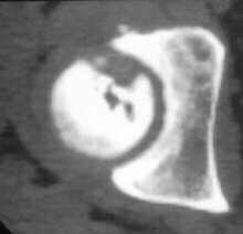
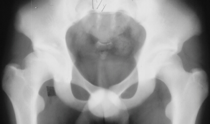
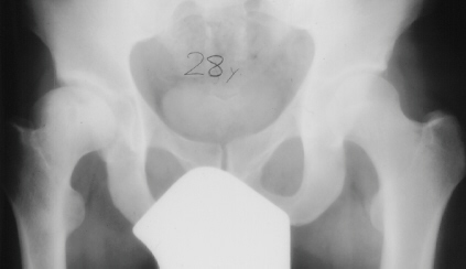
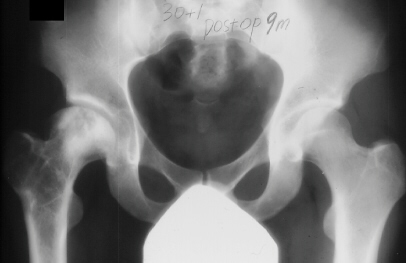
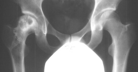
At 28 years of age, he had been having increasing pain in his right hip. He had a limp when he was tired. Positive Trendelenburg's sign was observed on the right. Internal rotation was limited to 25 degrees. All other motions were normal. X-ray showed cystic change. At the 29 years of age, he underwent bone graft of the right femoral head for degenerative cyst. At 35 years of age, only internal rotation was limited to 20 degrees. He still had pain in his hip that relates primarily to weather or to excessive walking. Only internal rotation was limited to 20 degrees. He had hip pain at the extremes of flexion and internal rotation. X-ray showed the progression of the cystic change. CT which was performed because of the question of loose bodies in the hip did not seem likely that the densities were actually in the hip joint. The antero-medial location of the cysts facing the anterior margin of the acetabulum was well depicted in CT.
28 yrs![]() ,
29 yrs
,
29 yrs![]() ,
30 yrs
,
30 yrs![]() ,
35 yrs
,
35 yrs![]()
![]()
 .
.
Snow S.W. et al. (A.I. duPont Institute) first reported four cases of this phenomenon in 1993. There were three boys and one girl. Age at onset ranged from 5+3 to 10 years (Table1. in the article).
Clinical features: