Brian J. McGinley, M.D., Resident, Orthopaedic Surgery
S. Jay Kumar, M.D., Attending, Orthopaedic Surgery
21 September 1995
ORTHOPAEDIC DEPARTMENT
THE ALFRED I. DUPONT INSTITUTE
WILMINGTON, DELAWARE
J. G. is a 14 3/12 y.o. boy who presented complaining of pain in his
right leg for 9 months and contracture of the right knee which has developed
over this period. There is no history of significant trauma. Born 3 weeks
premature with bilateral renal hypoplasia and a seizure disorder. Three
renal transplants have failed and he currently receives peritoneal dialysis
four times a day. Stunted growth and developmental delays during childhood
include short stature, standing at 16 months and walking at 2 years. He
sustained a right tibia and fibula fracture 6 years prior to presentation
after minimal trauma.
PHYSICAL EXAM: 5'4". Poor dentitia, right knee 15 degree flexion contracture, painful ROM of right hip and knee.
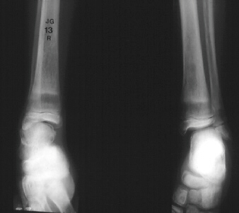
RADIOGRAPHIC EVALUATION: Osteopenia, subcortical resorption,
lucent metaphyseal bands, widening of metaphysis and "rugger jersey spine".
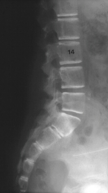
MEDICATION: Calcium carbonate and calcium acetate
Chronic renal failure often results in secondary hyperparathyroidism. The stimulus for elevated PTH is hypocalcemia, acidosis, diminished 1,25(OH)vitamin D and hyperphosphatemia.
Stunted growth and bony deformities including bowing of the lower extremities in ambulators. Rachitic rosary and prominent, widened inferior face due to mandibullar and maxillary overgrowth. Bone pain with or without fracture. Ectopic calcifications in the conjunctiva and skin. Slipped capital femoral epiphysis may also occur.
Early - Osteopenia
Thinning of cortices and trabeculae gives "ground glass" x-ray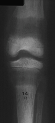
Salt and pepper skull
Physeal thickening and fraying of metaphysis, no cupping
Late epiphyseal ossification
Epiphyseal Slips - Preschool age: Proximal and distal femoral and distal tibial
Older Children: Proximal femoral and distal forearm
Late - Secondary hyperparathyroidism
Subperiosteal cortical resorption - Distal phalanx, end of clavicle, ischium, pubis, SI joints, metaphyseal-diaphyseal junction of long bones
Lucent metaphyseal bands - Growth zone changes are the best indicator of severity of secondary hyperparathyroidism
Bowing of long bones
Rugger Jersey spine - Osteosclerosis of the end plates of the vertebral bodies
Brown Tumor (rare) - Rib or jaw
Amyloidosis - Multiple bone cysts: Metacarpals, hip, wrist, prox humerus, pubic rami and proximal tibia.
GOALS:
PLAN:
TREATMENT:
Improved prognosis with dialysis and transplantation has led to prolonged life in these patients. Reversing the osteodystrophy with aggressive pharmacologic intervention can result in more normal growth and activity.