RICKETS IN AN ADOPTED CHILD
RICHARD B. ISLINGER, M.D., Orthopaedic Resident
WILLIAM G. MACKENZIE, M.D., Attending Pediatric Orthopaedic Surgeon
July 15,1996
CLINICAL CASE PRESENTATION
ORTHOPAEDIC DEPARTMENT
THE ALFRED I. DUPONT INSTITUTE
WILMINGTON, DELAWARE
CASE HISTORY:
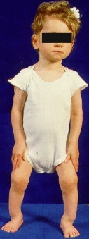
This patient was first seen here at the age of 2 and 1/2 years. She
was referred here with the diagnosis of nutritional rickets.
Significant past medical history included the following:
She was adopted from Russia and according the Russian medical documents
was born to a mother who suffered from alcohol abuse. She was born prematurely
(gestational age not available) at 2000gms, 44cm, with an apgar of 5/6.
Furthermore, she had previously been diagnosed with encephalopathy, anemia,
fetal alcohol syndrome and rickets. - Since arriving here in the U.S.,
the adopted mother had her on vitamin supplements for 7 weeks. The mother
states that the child began walking at the age of 27 months.

She was seen by both orthopaedics and pediatrics and her physical exam
was as follows:
- She was well below the 5th percentile for both height and weight
- There was delayed psycho-motor and speech development
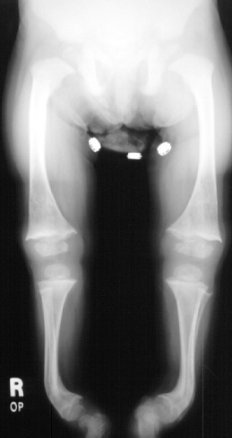
- Multiple rachitic deformities of the skeleton to include severe bowing
of the tibias bilaterally (45 degrees), pigeon chest (pectus carinatum),
thickened/widened wrists bilaterally and a trendelenberg gait on the left
side.
INITIAL LAB STUDIES:
Ca: 9.7 - nl
PO4: 6.2 - mildly increased
Alk Phos: 401 - mildly increased
UA: WNL
Vit D 1-25: 92 - mildly increased
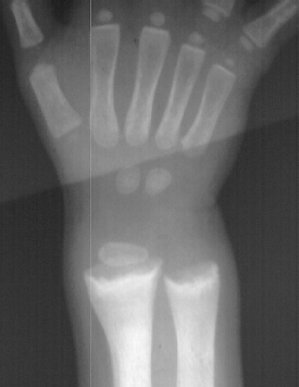
X-RAY STUDIES:
- "cupping" of the distal radius and distal femur
- widening of the physis
- angular deformities
She was diagnosed with nutritional rickets and was treated with Calciferol
1200ug\day. Her first follow-up was 5 weeks later where she was noted to
have a decreased Trendelenburg lurch and a decrease in her wrist thickening.
10 months later on follow-up she had a noticeable decrease in her tibial
bowing along with almost complete resolution of the growth plate abnormalities.
At her last follow-up on March 4th 1996 (she was 4 + 4yo) she had mild
anterior lateral bowing of her tibia bilaterally with a normal thigh-foot
angle and a mechanical axis that crossed the midline of her knee.
RICKETS:
Background:
- Earliest description of the disease - 1650
- Numerous etiological pathways but all involve a relative decrease in
Ca and/or PO4 which interferes with epiphyseal growth and normal
mineralization of the skeleton in the growing child.
- Despite the many possible causes, the clinical presentation, histology,
radiographic changes are virtually identical.
Clinical:
- Rachitic children are apathetic, irritable, with a height and weight
below the 3rd percentile
- Dentition is delayed, often they have severe caries and defective enamel
- Spine often has long, smooth dorsal kyphosis (rachitic catback) and
the chest shows enlargement of the costal cartilages (rachitic rosary)
- The extremities are most profoundly affected. Long bones are shortened
and deformed, ligamentous laxity is common, and fractures are common.
Histology:
- Thin cortices with thin and irregular trabeculae
- Widened osteoid seams (unmineralized segments of bone)
- Relatively normal resting and proliferative zone, with a grossly abnormal
zone of hypertrophy
- Zone of hypertrophy is widened 5-15 times normal
- Primary spongiosa show only limited bone formation
Radiographic:
- Osteopenia with thin cortices
- Physis is widened with irregular cupping
- Looser lines-transverse radiolucent lines on concave side of bone,
usually not extending to far cortex(most commonly found in renal osteodystrophy
and adult osteomalacia)
CAUSES:
- Deficiency- vitamin D intake is inadequate which causes diminished
absorption of Ca from the gut.
- Gastrointestinal- interference with bile salt production which
interferes with absorption of the fat soluble vitamin D. Ingested Ca forms
an insoluble soap with free fatty acids and is lost in the feces.
- Vitamin D-Resistant Rickets- four main types.
- Phosphate diabetes- vit D and Ca are normal but are hypophosphatemic
and cannot mineralize skeleton.
- Decrease in 1,25-dihydroxyvitamin D production- cannot convert 25-hydroxyvitamin
D to 1,25 form and thus cannot absorb Ca.
- End-organ insensitivity- gut cell is insensitve to 1,25 vit D
- Renal tubular acidosis- excretes excessive amounts of Ca
- Renal Osteodystrophy- Damage to the glomerulus causes retention
of PO4 while tubular injury reduces the production of 1,25 vit
D. Hyperphosphatemia further suppresses the production of 1,25 vit D andinhibits
renal reabsorption and GI absorption of Ca.. Renal osteodystrophy is characterized
by rickets, osteitis fibrosa (severe lysis of the skeleton due to secondary
hyperparathyroidism), osteosclerosis (20%),and ectopic calcification.
Management:
- Measurements of BUN, Creatinine, Ca, PO4, alk phos, 1,25
and 25 vit D, PTH, and urine Ca and PO4 to help establish the
diagnosis and categorize the type.
- First step is ALWAYS treat the underlying metabolic abnormality
first. Often after proper medical therapy the patient will go onto normal
growth and lifestyle.
- The limb malalignment is often dependent upon when the metabolic abnormality
occurred. If it is before two years old a varus deformity will occur, after
two years old a valgus deformity is most likely.
- If malalignment fails to improve after the underlying metabolic abnormality
is corrected, bracing and/or surgery may be indicated.
SELECTED LITERATURE REVIEW:
Normal development:
- The tibiofemoral angle in the newborn and infant is in varus (15 degrees).
At 18 to 24 months the tibiofemoral angle becomes more neutral. The tibiofemoral
angle then changes to valgus and is at its maximum at age three to four
(12 dgrees). It then corrects itself to that of the adult by age 7 (6 degrees
in boys, 7 in girls).
- Internal tibial torsion often accompanies physiologic genu varus and
accentuates the "bowlegs"
- Pes planus and external tibial torsion may accompany genu valgum- will
accentuate "knock knees"
Rachitic pathology
- Pathologic genu varum- large differential. Metabolic bone disease results
in bilateral pathology.
- Pathologic genu valgus- smaller differential. Renal Osteodystrophy
is the most frequent cause of bilateral pathologic valgus deformity
- Earlier surgical treatment for lower extremity abnormalities concentrated
on correcting the deformity by osteotomy and plating or multiple osteotomies
and intramedullary nailing. Ferris et. al., described a surgical procedure
in which he staged the osteotomies. The first stage consisted of multiple
diaphyseal osteotomies with "kebabing" over an intramedullary
nail, and this was performed at any age. They found correction of multidirectional
deformities with wedge osteotomies and plating difficult. Thesecond stage
consisted of a metaphyseal osteotomy which was helpful in correcting deformities
around the knees and was most successful if performed toward the end of
growth. Fixation was with a blade plate or staples
- Paley and Tetsworth performed corticotomies followed by placement of
external ring fixators with hinges placed at the apex of the deformities
for gradual correction. For multiapical deformities, they felt that meticulous
preoperative planning was the key for successful realignment of the extremity.
Their preoperative planning consisted of an elaborate method for elucidating
the mechanical axis deviation (MAD) of the extremity and the subsequent
amount of angular correction needed to realign themechanical axis
- Stanitski was successful treating rachitic limbs using the Ilizarov
technique. She found that decreasing the correction rate from 1 mm/day
down to 0.5mm/day in rachitic bone improved the quality of the regenerate
bone and she had no nonunions. With the Ilizarov technique, her patients
were allowed to weight bear as tolerated
When caring for rachitic deformities it is of paramount importance
to correct the underlying metabolic abnormality first prior to any surgical
intervention. If the metabolic abnormality is not corrected, surgical intervention
will likely fail.
TREATMENT RECOMMENDATIONS:
- Continued medical therapy as indicated
- Continued orthopaedic observation
- No surgical intervention needed at this time since the patient's deformities
continue to improve.
OUTCOME EXPECTATIONS:
- With the continued improvement seen in this child, we would expect
an excellent outcome in this individual
- If after the patient has reached or is near skeletal maturity and there
is limiting deformity still present, surgical intervention with expectations
of a good result can be undertaken.
REFERENCES:
- Ferris B, Walker C, Jackson A, Kirwan E.: The Orthopaedic Management
of Hypophosphatemic Rickets. J Pediatr Orthop 11:367-373; 1991.
- Kling TF Jr.: Angular Deformities of the Lower Limbs in Children. Orthop
Clin North Am 18:513-527; 1987.
- Paley D, Tetsworth K.: Mechanical Axis Deviation of the Lower Limbs.
Clin Orthop 280:65-71; 1992.
- Salenius P, Vankka E.: The Development of the Tibiofemoral Angle in
Children. J Bone Joint Surg (Am) 57:259-261; 1975.
- Stanitski DF.: Treatment of Deformity Secondary to Metabolic Bone Disease
With the Ilizarov Technique. Clin Orthop 301:38-41; 1994.
- Zaleske DJ. Metabolic and Endocrine Abnormalities. In: Lovell W, and
Winter RB, 4th ed. Pediatric Orthopaedics. Philadelphia: Lippincott-Raven,
1996.