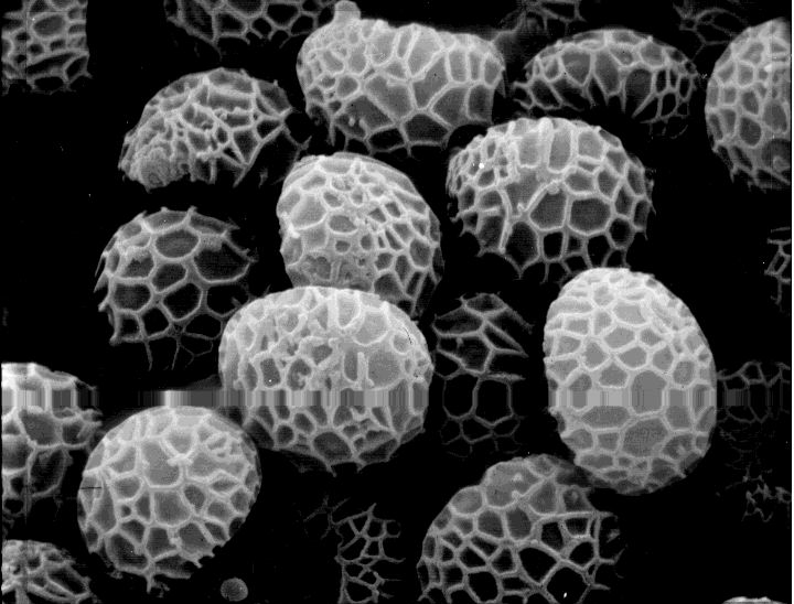
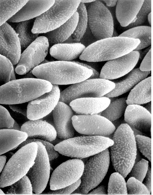
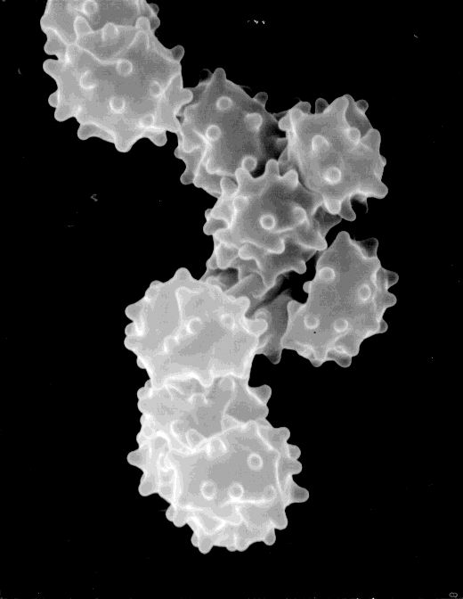
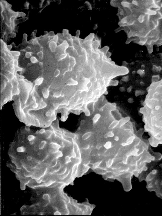
 Strobilomyces floccopus (U. New Hampshire)
 These images were kindly provided by Sam Ristich. (His annotations are included.)
The 3" x 5" grayscale prints were scanned in on a flatbed scanner, and I did a minimum of cropping and image manipulation before scaling the images for .gif and .jpeg compression and saving.
Click on the thumbnails to see a larger version.
Strobilomyces floccopus (U. New Hampshire)
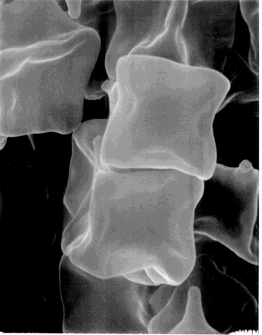
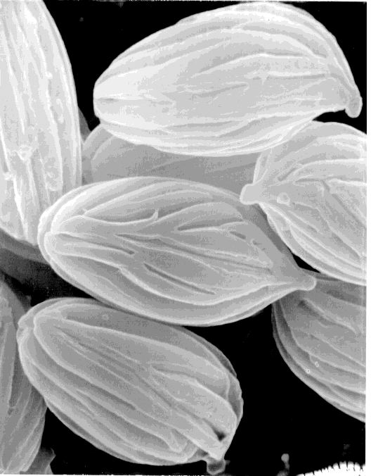
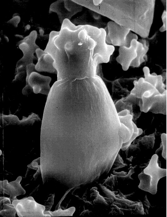
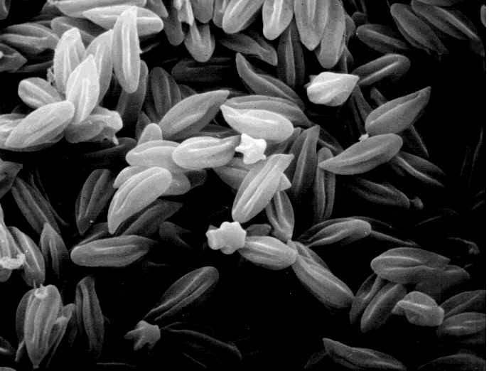 Clitopilus prunulus (U. New Hampshire)
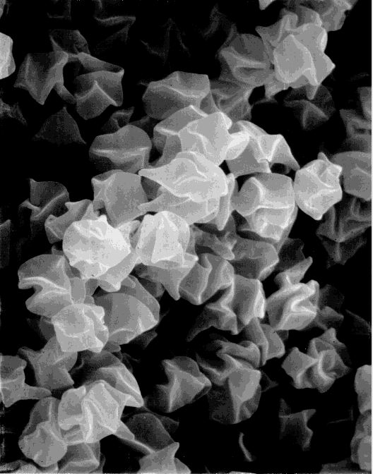
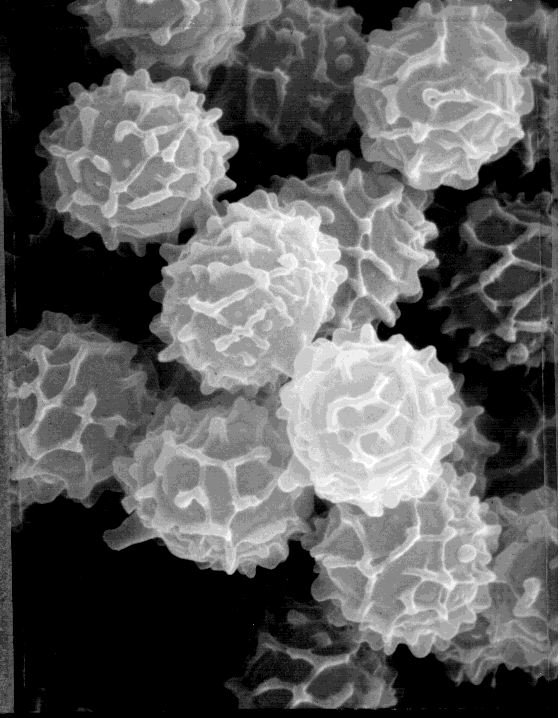
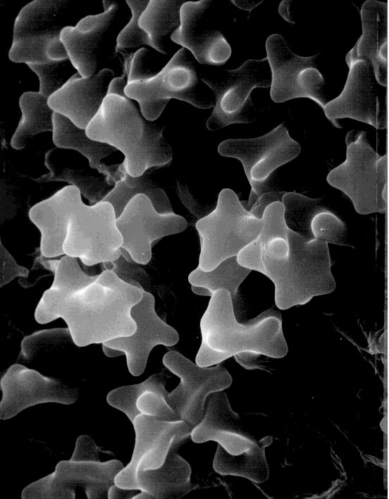
 Psathyralla velutina (1000 X on 3"x5") (large pore; grenade-like)
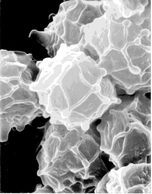
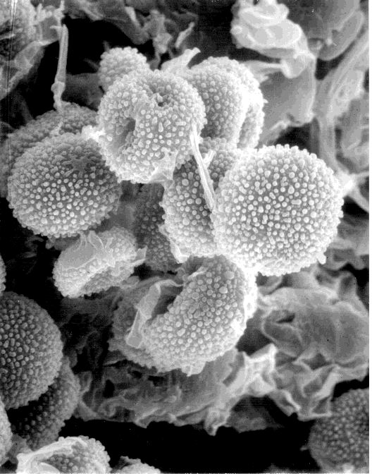 Sepedonium brunneum - an imperfect stage (1700 X)
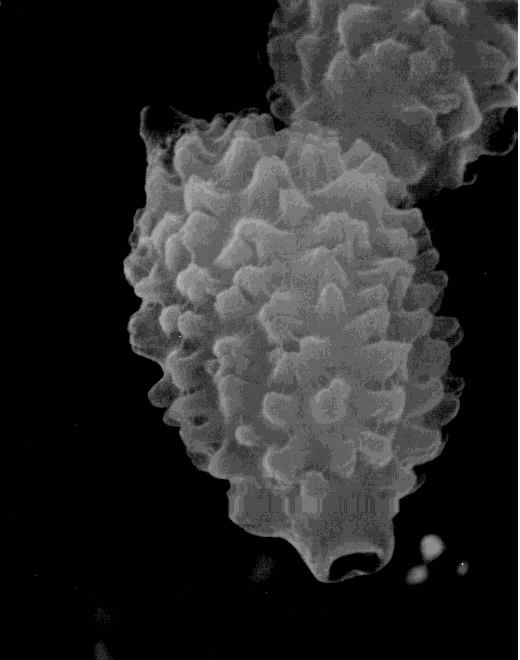
 Gyromitra korfii (1000 X)
 Entoloma salmoneum (5000 X)
 Entoloma sp. (many of these have collapsed) (2000 X)
 Lactarius volemus (Ithaca) (7500 X)
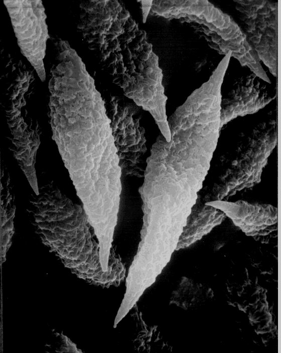
 Phellodon sp. (Ithaca) (6000 X)
 Boletus russellii (Ithaca) (5000 X)
 Hypomycetes lactifluorum (U. New Hampshire)
 Russula mariae (5000 X)
 Russula decolorans (5930 X)
 Inocybe sp. (cystidium)
 Inocybe sp.
copyright and all other rights reserved