 which progressed
 CHUN LI, M.D. Research Fellow, Orthopaedic Surgeon
ROBERT P. STANTON, M.D. Attending, Pediatric Orthopaedic Surgeon
May 20, 1996
CLINICAL CASE PRESENTATION
ORTHOPAEDIC DEPARTMENT
THE ALFRED I. DUPONT INSTITUTE
WILMINGTON, DELAWARE
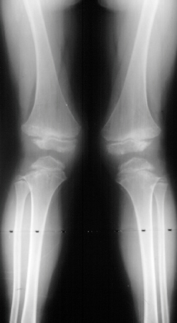
This patient is an 11 year old white male with a diagnosis of Multiple
Epiphyseal Dysplasia (MED). Initially he presented with x-rays revealing
mild genu valgum
which progressed ![]() such that he required surgery. He had supracodylar osteotomy which was
performed in 1993 for genu valgum
such that he required surgery. He had supracodylar osteotomy which was
performed in 1993 for genu valgum ![]() .
He was last seen in August 1995 for low back pain for which he was prescribed
stretching exercises. Since the last visit he has had no problems with
his back and denies any complaints in his lower extremities, with the knee
valgus remaining stable
.
He was last seen in August 1995 for low back pain for which he was prescribed
stretching exercises. Since the last visit he has had no problems with
his back and denies any complaints in his lower extremities, with the knee
valgus remaining stable ![]() .
.
Examination revealed a significantly obese boy in no acute distress. Examination of his back showed no evidence of any curvature. There was no tenderness of his entire spine. Examination of his legs revealed bilateral genu valgum with an intermalleolar distance of 13.5 cm. There was no evidence of any knee effusion or tenderness of the knees. Neurovascular status of the lower extremities was normal with 5/5 muscle tone bilaterally.
MED first was described by Fairbanks in 1937. Inherited disorder, that is predominantly autosomal dominant. It commonly begins with painful or stiff lower extremities. Hip and knee joints are most frequently affected. The affection occurs symmetrically in most cases.
The basic defect is a disturbance in the development of the epiphyseal ossification centers. Enchondral ossification is disorganized, and epiphyseal cartilage cells are irregular with disordered columns and areas of degeneration.
Lower extremity joint pain and stiffness, short stature, short stubby hands and feet along with a waddle gait are features of MED. Patients with MED usually have normal intelligence. Child patients are delay in walking. Adult patients' height usually range from 145 to 170 cm.
In radiograms, the principal findings are the delay and irregularity of ossification of the epiphyses, which are markedly fragmented and mottled. They appear flattened.
In differentiating multiple epiphyseal dysplasia from juvenile osteochondritis, including Perthes' disease, a point to remember is that multiple epiphyseal dysplasia is multiple and bilateral. A single joint of Perthes' disease may appear indistinguishable from that of epiphyseal dysplasia, but skeletal survey will reveal the difference. Osteochondritis is also an acquire rather than a familial disorder and usually is painful and becomes worse with observation if no treatment is given. Osteochondritis occurs in a previously normal epiphysis, while in dysplasia Epiphysialis multiplex the epiphysis is abnormal and ossifies irregularly.
1. Weight control should be emphasized early on.
2. Patients should be refrained from contact sports with high impact activities.
3. Osteotomies about the knee can be done in order to correct alignment.
4. Patients will most likely require joint replacement surgery in adulthood.