BILATERAL SLIPPED CAPITAL FEMORAL EPIPHYSIS
CHARLES J. ODGERS IV, M.D., Resident, Orthopaedic Surgery
KIRK DABNEY, M.D., Attending Pediatric Orthopaedic Surgeon
March 19, 1996
CLINICAL CASE PRESENTATION
ORTHOPAEDIC DEPARTMENT
THE ALFRED I. DUPONT INSTITUTE
WILMINGTON, DELAWARE
CASE HISTORY
An 11-year-old boy presented with a one year history of right knee pain.
He stated that his knee recently "gave way" when running down
the stairs at school. He described his discomfort as a deep achy pain on
the medial aspect of his knee. There was no history of trauma, nor had
the pain limited his activities until recently. Pt denied numbness, paresthesias,
or weakness in his right leg. He denied fevers, chills, and any recent
illnesses. He denied history of hip or groin pain.
PHYSICAL EXAM
Examination revealed an obese adolescent male in no acute distress.
Height- 149cm(50%) Weight- 59 Kg(>95%). Right knee appeared atraumatic
without any areas of point tenderness. There was diffuse pain in the knee
with passive ROM. Examination of the hips revealed no tenderness, however
there was increased ER and decreased IR of the right hip compared to the
left. There were no gait abnormalities.
RADIOGRAPHS
An AP pelvis and frog-leg lateral radiographs of both hips were obtained.
On the AP pelvis radiograph, there was notable widening and irregularity
of the proximal femoral epiphyseal growth plate, and Klein's line did not
intersect the epiphysis .
The frog-leg lateral view of the right hip demonstrated an obvious grade
1 slipped capital femoral epiphysis
.
The frog-leg lateral view of the right hip demonstrated an obvious grade
1 slipped capital femoral epiphysis .
.
FOLLOW-UP
The patient was admitted to the hospital, placed on strict bedrest,
and he had an in situ pinning of his right hip the next day. There were
no postoperative complications, and he was discharged one day later. By
the time of his follow-up visit three weeks later, he was riding his bike
and had discarded his crutches. An AP pelvis  and frog-leg lateral radiographs of both hips were obtained which were
normal. At his three-month follow-up visit, the patient complained of a
two week history of vague left hip pain. AP
and frog-leg lateral radiographs of both hips were obtained which were
normal. At his three-month follow-up visit, the patient complained of a
two week history of vague left hip pain. AP  and frog-leg lateral radiographs revealed a grade 1 left slipped capital
femoral epiphysis. He had an in situ pinning of his left hip the next day
without complications
and frog-leg lateral radiographs revealed a grade 1 left slipped capital
femoral epiphysis. He had an in situ pinning of his left hip the next day
without complications  .
.
DISCUSSION
SCFE- Clinical Presentation
- History- duration of Sx
- Symptoms nonspecific- delay in Dx
- Groin, medial thigh, or knee pain
- Limitation of ROM- esp. IR
- Antalgic limp, abductor lurch
- Always examine other hip
SCFE- Radiographic Presentation
- Radiographic assessment- need AP and lateral views(most important)
- True lateral view better than frog-leg
- Klein's line does not intersect epiphysis on AP view
- Widening and irregularity of physis
- Height of epiphysis decreased in central acetabulum on AP view- epiphysis
has slipped posteriorly
Epidemiology of Bilateral SCFE
- Incidence of B/L SCFE ~ 25%(21-37%)- 50% present simultaneously
- Long term studies - identified changes c/w bilateral involvement in
60-80% of pts with known unilateral SCFE( silent slips)
- Incidence of B/L involvement is increased in males, African-Americans,
obese pts, and pts with a young age(<13.5yrs. for boys and <11.5
in girls) at initial presentation
- When B/L SCFE occurs sequentially, second slip presents within 18 months
of initial slip in 88% of patients
Bilateral SCFE and Endocrine Disorders
- Bilateral SCFE associated with an endocrine disorder ~ 7% of patients
- Hypothyroidism- most common
- Wells and coworkers-prevalence of bilaterality in SCFE associated with
endocrine disorders was eventually 100%
Prophylactic Pinning of the Contralateral Hip in Patients with SCFE
- Proponents- Emphasize rate of B/L disease and higher risk of osteoarthritis
with the increase in slip severity
- Opponents- Stress that in situ pinning can be associated with severe
complications and that many hips would be treated unnecessarily
- Trend- Observation of the unaffected hip
- Patient should be followed with serial radiographs(AP pelvis and lateral
view of hip) every 3-4 months
- Make pt aware of prodromal symptoms
- If symptoms develop-return immediately for evaluation
- 1. Patient with known metabolic or endocrinologic disorder
- 2. Patient with inability to obtain timely and appropriate follow-up
due to personal or family circumstances
REFERENCES
- Emery R, Todd R, Dunn D: Prophylactic pinning in slipped upper femoral
epiphysis: Prevention of complications. J Bone Joint Surg Br 1990; 72:217-219.
- Hagglund G, Hansson L, Ordeburg G, Sandstrom S: Bilaterality in slipped
upper femoral epiphysis. J Bone Joint Surg Br 1988; 70:179.
- Klein A, Joplin R, Reidy J, Hanelin J:Roentgenographic features of
slipped capital femoral epiphysis. Am J Radiology 1951; 66:361.
- Loder R, Aronson D, Greenfield M: The epidemiology of bilateral slipped
capital femoral epiphysis: A study of children in Michigan. J Bone Joint
Surg AM 1993; 75:1141-1147.
- O'Beirne J, McLoughlin R, Dowling F, et al: Internal fixation using
single central pins. J Pediatr Orthop 1989; 9:304-307.
- Rappaport E, Fife D: Slipped capital femoral epiphysis in growth hormone
deficient patients. Am J Dis Child 1985; 139:396-399.
- Segal L, Davidson R, Robertson W Jr, et al: Growth disturbances of
the proximal femur after pinning of juvenile slipped capital femoral epiphysis.
J Pediatr Orthop 1991; 11:631-637.
- Ward W, Stefko J, Wood K,, et al: Fixation with a single screw for
slipped capital femoral epiphysis. J Bone Joint Surg Am 1992; 74:799-809.
- Wells D, King J, Roe T, et al: Review of slipped capital femoral epiphysis
associated with endocrine disease. J Pediatr Orthop 1993; 13:610-614.
 and frog-leg lateral radiographs of both hips were obtained which were
normal. At his three-month follow-up visit, the patient complained of a
two week history of vague left hip pain. AP
and frog-leg lateral radiographs of both hips were obtained which were
normal. At his three-month follow-up visit, the patient complained of a
two week history of vague left hip pain. AP  and frog-leg lateral radiographs revealed a grade 1 left slipped capital
femoral epiphysis. He had an in situ pinning of his left hip the next day
without complications
and frog-leg lateral radiographs revealed a grade 1 left slipped capital
femoral epiphysis. He had an in situ pinning of his left hip the next day
without complications 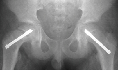 .
.