
JOHN ERGENER, M.D., Resident, Orthopaedic Surgery
WILLIAM G. MACKENZIE, M.D. Attending Pediatric Orthopaedic Surgeon
CLINICAL CASE PRESENTATION
ORTHOPAEDIC DEPARTMENT
THE ALFRED I. DUPONT INSTITUTE
WILMINGTON, DELAWARE
The patient is an 8 year and 7 month old while male child who presented for orthopaedic evaluation after radiographs were taken of his left knee. The child reported that he injured his left knee about two weeks ago when he had fallen of his bicycle. He had been seen by his primary care physician when his pain and swelling persisted. The child complained of pain and swelling and difficulty moving his left knee. He has been able to bear weight and ambulate although with a limp.
The patient was able to ambulate with an antalgic gait and the left knee held in mild flexion. The left knee lacked full range of motion. Extension was approximately 10 degrees short of full. Flexion was to approximately 120 degrees, about 20 degrees less than the opposite side.
There was a moderate effusion evident by blotting of the patella. There were no specific areas of tenderness although ranging the knee was bothersome with mild apprehension. No ligamentous instability was noted during the exam. Exams of the hip and ankle were normal bilaterally as was the right knee.
AP, lateral and obliques of the left knee showed an effusion with multiple
radiodense bodies that were felt to be intraartictilar
![]() .
No fractures or dislocations were evident. Comparison views of the right
knee were unremarkable.
.
No fractures or dislocations were evident. Comparison views of the right
knee were unremarkable.
Synovial Chondromatosis
Arthroscopic removal of loose bodies with probable extensive synovectomy and possible arthrotomy.
Arthroscopic removal of loose bodies with probable extensive synovectomy. This 8 + 7 year old male child underwent left knee arthroscopy with removal of loose bodies and extensive synovectomy. Posteromedial and posterolateral portals as well as standard anteromedial and anterolateral portals were used. Multiple loose bodies were found on the order of 15- 25. They measured approximately 4-6mm x 3-4mm x 3-4mm. They resembled the so called rice bodies. Synovial reaction was also evident in the areas where these loose bodies seemed to be developing. The articular surfaces appeared to be affected with grades I - II chondromalacia. The menisci and ligaments were in good condition.
Post-operatively the left knee was held in an immobilizer. The patient
was allowed to weight bear as tolerated with crutches. Over the course
of two weeks the crutches and immobilizer were weaned. At three weeks post-operative
the patient was with mild residual effusion but without pain. Range of
motion was improved with the ability to reach full extension. The patient
is doing well in the early post-operative period 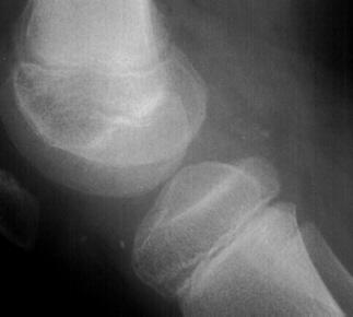 .
.
(Also called synovial osteochondromatosis, synovial chondrometaplasia, synovial chondrosis, synovial chondromata)
Synovial chondromatosis is a rare benign condition that involves the synovial lining of joints, bursae or tendon sheaths. It is characterized by the development of multiple osteochondral loose bodies. Joints are the most commonly affected. It is a monoarticular condition. In order of frequency is the knee, elbow, shoulder and hip. It has also been reported in the temporomandibular joint. It is a process whereby the synovium undergoes metaplasia and ultimately forms cartilage loose bodies. These loose bodies may go on to ossify. This has been described by Milgram to occur in three stages. First, the synovium (for unknown reasons) undergoes metaplasia. This is an active process of intrasynovial proliferation of cartilage nests. At this time cartilage nodules are formed that remain attached to the synovium. In the second stage, the nodules become detached and are present as loose bodies. It is felt that patients may become symptomatic at this time. In the third phase, the synovium has burnt itself out. There is no longer evidence of metaplasia but there is still loose bodies present. These may have become ossified by this time.
The etiology of synovial chondromatosis is not known. Some suspect that it is due to synovial irritation. This may be from trauma or possibly from infection, but this is unclear. No organism has ever been cultured. No significant correlation has been made with trauma.
This condition is rare and few reports are made of patients in the pediatric population. This 8 year old is the youngest of my literature review. This disease is reported to be more common in males and usually presents in the ' )rd to 5th decades. It is difficult to judge the onset but it is believed to be insidious over 6 months to years.
A patient will usually complain of swelling of the afflicted joint. There may be a dull ache present. Patients may also have symptoms of locking, stiffness, limitation of joint motion, crepitus or giving way. Complaints are often tied to a traumatic event but this is difficult to confirm.
On physical exam, an effusion may be present. There may be tenderness of the joint or pain on range of motion. The range of motion may be limited. A palpable tender mass may be present.
Evidence on imaging studies depends on the stage of disease. Until the loose bodies are ossified or calcified they may be radiographically invisible. This often leads to an unfortunate delay in treatment.
Although the metaplastic changes of the synovium are reported to be self limited, damage to the joint from multiple loose bodies may lead to early degenerative arthritis. Needless to say the pain and other symptoms will be relieved by evacuation of the loose bodies. With the advent of arthroscopy this has become a relatively easy and less invasive of a procedure. Joints other than the knee, may still lend themselves to arthrotomy and exploration. Controversy exists as to whether a synovectomy should be performed. Recently, from a retrospective study of 13 patients with synovial chondromatosis of the knee, an argument has been made to perform a thorough synovectomy. Of the five patients that had removal of loose bodies alone, three had recurrence of loose body formation (Ogilvie-Harris, 1994). None of the eight treated with initial removal of loose bodies and a thorough synovectomy had recurrence.
The offending entity is the synovium which may appear hyperemic and with thickening and fronds. Cartilaginous bodies are initially enveloped and then break free to form loose bodies. They are small grey-white shiny polyploid bodies. Many of the loose bodies are sequestered in the posterior aspect of the knee. This makes total evacuation difficult.
Recent immunohistological studies have been done to indeed show that these cartilaginous loose bodies are the result of metaplasia of the synovium (Apte, 1992). The histologic appearance of this process shows well-defined cartilage nests embedded in a layer of synovium. There may significant pleomorphism. There may be evidence of advancing stages with maturation of the cartilage with mineralization and ossification. Mitotic figures are not a prominent feature of the proliferating chondrocytes. There may, however, be some plump hyperchromatic nuclei.
Synovial chondromatosis is reported to be a benign self-limiting disease process. Chondrosarcoma of synovium has been described. There is controversy as to whether malignant degeneration takes place leading to a secondary malignant process. However, there have been at least 19 well documented cases of such a secondary malignant process. Unfortunately, there are no clear cut radiological studies that can differentiate the entities.
Histologically the malignant tumor can be differentiated by its loss of a solid matrix, a so called "runny" matrix, hypercellularity with crowding and spindling of the nucleus and necrosis or local invasion. Metastasis is the hallmark of malignancy and pulmonary spread was present in 7 of the 19 cases.
Synovial chondromatosis is a disease process of the synovium, whereby metaplasia takes place to form cartilaginous loose bodies. Symptoms develop form the irritation of the synovium and mechanical properties of the loose bodies. The process is insidious and may go on for years before being diagnosed. Once diagnosed removal of the loose bodies with synovectomy provides the best chance for eradication of the disease.
In the presented case of this 8 year old boy, close follow up is necessary to diagnose recurrence and hopefully minimize the risk of degenerative arthritis.