Module 35, Pediatric Orthopaedist Level
 1,
1,
 2,
2,
 3,
3,
 4,
4,
 5,
5,
 6,
6,
 7
7
A 2 year and 6 month old white male presented to clinic with a 2-3
month history of limping on the right lower extremity. There was no history
of trauma to the extremity, or a history of constitutional symptoms such
as fever, chills, sweats, malaise or weight loss. Prenatal, perinatal and
postnatal course were all normal. Examination of the right lower extremity
was significant for a distinct absence of ankle dorsiflexion and eversion
activity to motor testing. Plantar flexion strength was graded as 5/5.
Sensation was intact on the entire plantar aspect of the foot, with patchy
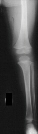
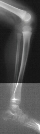
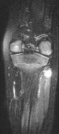
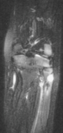
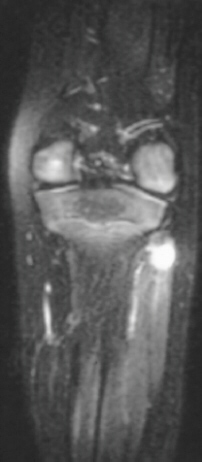
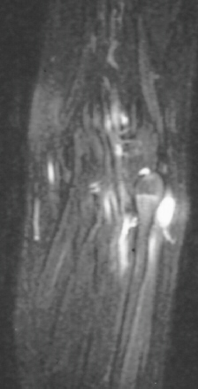
sensation on the dorsum of the foot, including the first web space. Radiographic
examination of the left knee revealed no osseous abnormalities (images
1 and 2). A fullness of the soft tissues was evident over the fibular head.
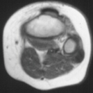
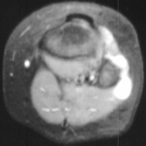
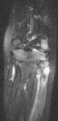
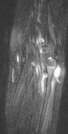
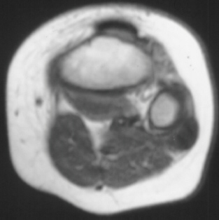
MRI examination of the left knee displayed a 2 cm. by 0.8 cm. lobulated
mass in the region of the common peroneal nerve with no enhancement (images
3and 4). On the T-2 weighted sequences there was abnormal increased signal
of the tibialis anterior, extensor digitorum longus and peroneus longus
muscle bellies suggesting atrophy (images 5, 6 and 7).
Question 35A

 Electrodiagnostic
tests of the peroneal nerve would be expected to show a definite non-functioning
nerve.
Electrodiagnostic
tests of the peroneal nerve would be expected to show a definite non-functioning
nerve.
Question 35B
Based on this imaging, the differential diagnosis should include all the
following: Neurilemmoma, 2) Neurofibroma, 3) Ganglion, 4) Hemangioma, 5)
Neuroma.
Question 35C
Based
on the above information you would do a biopsy of the most prominent part
of the mass and wait for permanent microscopy results.
Question 35D
Surgical
exploration revealed a lobulated mass compressing and encompassing the
peroneal nerve. The stalk of the cyst was found to be originating from
the proximal tibio-fibular joint. This sounds like a benign joint ganglion
and you would inform the parent that you expect the nerve to return to
full function.
Question 35E
The
following is a result of the biopsy. .
This is most likely a Neurilemmoma.
.
This is most likely a Neurilemmoma.
Go to Next Question Module, Go
to General Orthopaedist Level Modules, Go
to Question Module Home Page, Go
to Case Presentation Home Page, Orthopaedic
Department Home Page.
 3,
3,
 4,
4,
 5,
5,
 6,
6,
 7
7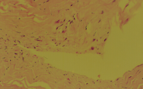 .
This is most likely a Neurilemmoma.
.
This is most likely a Neurilemmoma.