


TEST Passive ROM Strength Spasticity Motor Control Comments
                     Rt    LT    Rt   LT   Rt   LT   Rt    LT
Hip Flexion          120   115   4    5    1    1    yes   yes
hip abduction (EXT   25    22    4    3    4    0    yes   yes
KN))
Hip extension        8     0     5    5              yes   yes
Hip Int Rotation     40    55
(EXT)
Hip ext Rotation     25    15
(EXT)
Thomas test          23    18
Knee extension       -9    5     5    5    4    3    yes   yes   Rectus spasticity
Knee Flexion         135   145   5    4    1    2    yes   yes
Knee reflex                                2    3
Popliteal angle      70    70
Ely Test             85    85
Ankle Dorsiflexion   8     0                                     With knee Flexion
Ankle dorsiflexion   3     -10   5    4              yes   no    With Knee
                                                                 Extension'
Ankle Plantar        35    50    4    5    3    1    yes   yes
flexion
Foot inversion       45    40    5    5    3    3    yes   yes   Tibialis
                                                                 Posterior
Foot eversion        33    -5    5    5              yes   yes   Perineals
Thigh Foot angle     e-35  e-10                                  done with knee
                                                                 flexed
Babinski                                             no    no
Clonus                                     5    12
                                           bts  bts
Standing GMFM                                                    32 of 36
                                                                 possible points
Leg length measured  87.5  87                                    Measured supine
The major findings on the kinematic were slow speed, normal cadence, and symmetrical but short step lengths.
Characteristic:                        Trial        Normal       % Normal

TEMPORAL CHARACTERISTICS:
Velocity, cm / sec                      91.3        120.0           76.1
Cadence, steps / min                   118.0        122.0           96.7
Stride Time, sec                         1.0          1.0          101.7
SPATIAL CHARACTERISTICS:
Stride Length, cm                       92.8        119.0           78.0
Right Step Length, cm                   47.7         59.4           80.3
Left Step Length, cm                    45.1         59.4           76.0
Step Width, cm                          17.1         12.0          142.7
Pelvic Width / Ankle Spread Ratio       0.98          2.0           48.9
The pelvis was rotated leading with the right forward 5 to 30 degrees
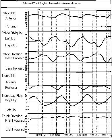 .
The left hip was adducted 5 to 25 degrees and internally rotated 20 degrees.
the right was externally rotated 10 degrees
.
The left hip was adducted 5 to 25 degrees and internally rotated 20 degrees.
the right was externally rotated 10 degrees 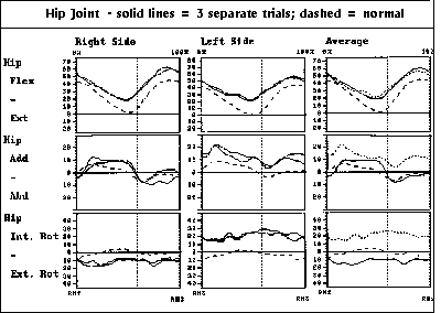 .
The right knee had increased mid-stance flexion of 30 degrees and good
flexion in swing. The left had flatted and decreased swing flexion and
both had increased knee flexion at foot contact at 35-40 degrees. The right
tibia demonstrated valgus of 25 degrees and external rotation of 20 degrees.
Dorsiflexion of the right ankle was good but plantar flexion was limited
to neutral. The left had limited dorsiflexion coming only to neutral. Foot
progression on the right was external 20-30 degrees and on the left was
internal 5 degrees
.
The right knee had increased mid-stance flexion of 30 degrees and good
flexion in swing. The left had flatted and decreased swing flexion and
both had increased knee flexion at foot contact at 35-40 degrees. The right
tibia demonstrated valgus of 25 degrees and external rotation of 20 degrees.
Dorsiflexion of the right ankle was good but plantar flexion was limited
to neutral. The left had limited dorsiflexion coming only to neutral. Foot
progression on the right was external 20-30 degrees and on the left was
internal 5 degrees 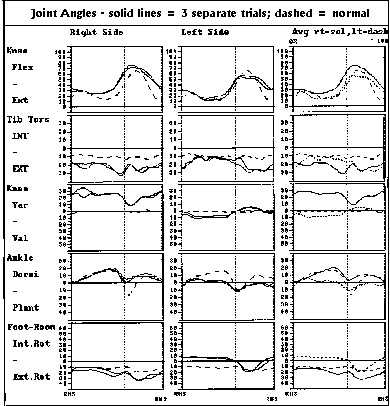 .
.
The recording of the Rectus muscle shows significant swing phase activity
bilaterally and very minimal stance activity. Both hamstring muscles show
significant constant activity but do have additional activity early stance
phase![]() . Tibialis anterior muscle has predominantly early swing phase activity
with significant underlying constant activity. The right gastrocnemius
has predominantly stance activity with good late stance phase contraction.
The left gastrocnemius has no recognizable pattern
. Tibialis anterior muscle has predominantly early swing phase activity
with significant underlying constant activity. The right gastrocnemius
has predominantly stance activity with good late stance phase contraction.
The left gastrocnemius has no recognizable pattern ![]() .
.
![]()
![]() A
derotation osteotomy of the left femur is definately indicated.
A
derotation osteotomy of the left femur is definately indicated.
![]()
![]() The
right tibia has 35 degrees of external torsion which is a major cause of
the knee valgus. Both of these can be corrected through a proximal tibial
osteotomy.
The
right tibia has 35 degrees of external torsion which is a major cause of
the knee valgus. Both of these can be corrected through a proximal tibial
osteotomy.
![]()
![]() One
option would be to do a external rotation femoral osteotomy on the left
and a proximal tibial osteotmy on the right. It would be important to do
the femur first.
One
option would be to do a external rotation femoral osteotomy on the left
and a proximal tibial osteotmy on the right. It would be important to do
the femur first.
![]()
![]() Reconstruction
of the left hip to gain good reduction and acetabular coverage will require
a pelvic osteotomy like a Dega. The indication for reconstructing this
hip remain unclear.
Reconstruction
of the left hip to gain good reduction and acetabular coverage will require
a pelvic osteotomy like a Dega. The indication for reconstructing this
hip remain unclear.
![]()
![]() Based
on the data above this boy needs a tnedon achilles lengthening on the left
in addition to the right knee and left hip procedure. This is all that
will be needed at this surgical event.
Based
on the data above this boy needs a tnedon achilles lengthening on the left
in addition to the right knee and left hip procedure. This is all that
will be needed at this surgical event.
Go to General Orthopaedist Level Modules, Go to Question Module Home Page, Go to Case Presentation Home Page, Orthopaedic Department Home Page.